flow cytometry results explained
The detected of anti-platelet. Get reliable results you can trust with our range of KO validated antibodies.

Overview Of High Dimensional Flow Cytometry Data Analysis A Fcs Download Scientific Diagram
They may order a flow cytometry test to see if the lymphocytes are clonal seen in.

. Flow cytometry intracellular staining protocol. Summary of the Assay Qualification Process for Flow Cytometry Assays. Blot results will be influenced if the target epitope is destroyed.
Accuracy of Urine Flow Cytometry and Urine Test Strip in Predicting Relevant Bacteriuria in Different Patient Populations. Infectious causes of lymphocytosis include. Flow cytometry data were analyzed using FloJo 932 Tree Star Inc.
Flow Cytometry Results Interpretation Seurat and the Cell Sorting Impressionist. BoatUS Magazine official publication of the Boat Owners Association of The United States BoatUS provides recreational boating skills DIY maintenance safety news lifestyle and personality profiles and insight from top expertsThe award-winning boating magazine publishes several. Because cells have negative intracellular potentials the electrical force.
Someone could be a frequent drug user and stop using drugs a month or two before a drug test and receive a negative test. B16F10 tumors harvested from mice implanted with hydrogels or email protected were analyzed by flow cytometry 2 days after treatment. During the past decade or two the field of biomarkers short for biological markers has witnessed immense development.
Thus they have higher specificity resulting in lower background. From BoatUS Magazine Americas Most-Trusted Boating Magazine. Available autologous transduced T cells with greater than or equal to 20 expression of GPC3 CAR determined by flow-cytometry and killing of GPC3-positive targets greater than or equal to 20 in cytotoxicity assay.
In addition too much antibody may result in a. The complexity of the field mirrors its multi-faceted applications and it is easy to get. Whole-body computed tomography and flow cytometry in peripheral blood should be considered.
Overall the results in all analyzed samples underline the power of this 13-parameter flow cytometric SST strategy to reliably confirm or exclude lymphoma localization. If youve never seen flow cytometry results before your first guess might be that the ghost of Seurat lives inside your machine. Obtain continuous cell assay data points over the course of the entire experiment.
The membrane potential and the differences in ion concentrations between the intracellular and the extracellular spaces. Direct flow cytometry protocol. Chemical structures were drawn using ChemDraw Ultra Cambridgesoft.
A Representative flow cytometric analysis of T cell infiltration within the tumor and B corresponding quantification results. Lymphocytosis often results from viral infections. Beginning with the underlying mathematical component the method behind the madness.
Directly conjugated neural markers. Results in E and F are mean SD from one of three representative experiments. Blot results will be stable even though a few epitopes are.
With 20 markers the doctor would already have to compare about 150 two-dimensional images he said thats why its usually too costly to thoroughly sift through the entire data set The team explored how AI could be used to carry out flow cytometry testing. The appropriate controls for this multicolor flow cytometry experiment include an unstained sample to look at autofluorescence single stained samples to allow compensation data to be generated isotype controls to check for non-specific background staining and FMO controls to account for spreading of the data. This makes false-negative results possible.
Typically the validation of a flow cytometry panel for GLP or GcLP applications includes test scripts to assay the following characteristics. The study investigators explained in their report. BMC Infectious.
Informed consent explained to understood by and signed by patientguardian. Precision includes repeatability and reproducibility. Simple label-free protocol measures cell killing continuously from seconds to days making it ideal for immunotherapy applications.
Bacteria and parasites can also cause infection resulting in a high lymphocyte count. 1 Serum protein electrophoresis and quantification of immunoglobulin classes must be performed for all. Flow Cytometry Basics Guide.
Dr Krawitz explained that sample analysis using flow cytometry is very time-consuming. Gehringer Christian et al. General techniques will be explained.
They recently described the assay and its use in a study with results reported in the journal Blood Advances. Monitor cell behavior in real time in a 6 x 96-well format on the xCELLigence RTCA MP. Polyclonal antibodies recognize more epitopes and they often have higher affinity.
Chapter 6 Basic Rules for Building Multicolor Panels. Antibody will bind at low affinity and create background that will reduce the resolution and therefore cloud your results. Flow cytometry analysis indicated that whereas SIINFEKL resulted in the expected antibody recognition at the cell surface of untreated cells the SIINwEKL peptide did not Fig.
Cells and Model Organisms. Researchers have developed a flow cytometry-based method to assist with diagnosis of vaccine-induced immune thrombotic thrombocytopenia VITT. All these cases 1313.
This test shows if you have a higher-than-normal amount of lymphocytes. Published July 9 2016. Monoclonal antibodies recognize single specific antigenic epitope.
And Research Applications Explained. An excitable membrane has a stable potential when there is no net ion current flowing across the membrane. Patientguardian given copy of informed consent.
Two factors determine the net flow of ions across an open ionic channel. Ferroptosis cannot be explained by a simple increase in H 2 O 2-dependent iron-catalyzed ROS production ie.
Blog Flow Cytometry Data Analysis I What Different Plots Can Tell You
Veterinary Sciences Free Full Text Flow Cytometry Detected Immunological Markers And On Farm Recorded Parameters In Composite Cow Milk As Related To Udder Health Status Html

Show Dot Blot Analysis Of Flow Cytometry Data Of Cd4 Cd8 Of Two Cases Download Scientific Diagram
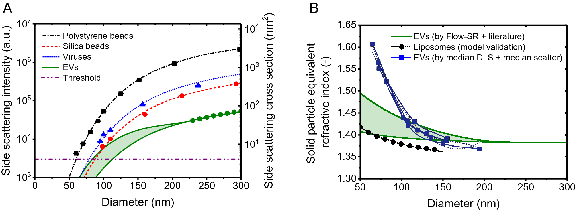
Misinterpretation Of Solid Sphere Equivalent Refractive Index Measurements And Smallest Detectable Diameters Of Extracellular Vesicles By Flow Cytometry Scientific Reports
Chapter 4 Data Analysis Flow Cytometry A Basic Introduction
Blog Flow Cytometry Data Analysis I What Different Plots Can Tell You

Flow Cytometry Basics Flow Cytometry Miltenyi Biotec Technologies Macs Handbook Resources Miltenyi Biotec Ireland

Overview Of High Dimensional Flow Cytometry Data Analysis A Fcs Download Scientific Diagram
Representative Flow Cytometry Data A Negative Control Cells Download Scientific Diagram

How To Identify Bad Flow Cytometry Data Bad Data Part 1 Cytometry And Antibody Technology

Typical Data From A Two Color Flow Cytometry Experiment To Measure Cell Download Scientific Diagram

What Is Flow Cytometry Facs Analysis

Flow Cytometry Tutorial Flow Cytometry Data Analysis Flow Cytometry Gating Youtube
Quantitative Flow Cytometry Measurements Nist

Flow Cytometry Control And Standardization Beads
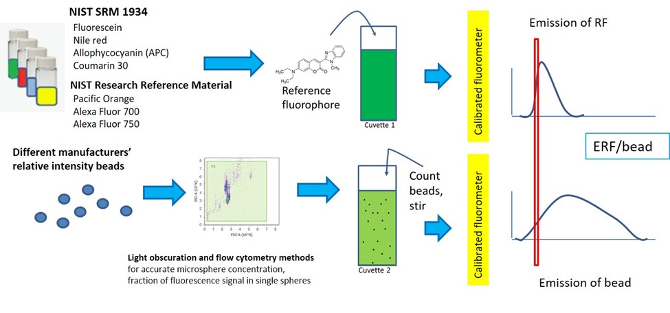
Quantitative Flow Cytometry Measurements Nist
Flow Cytometry Based Platelet Activation Markers And State Of Inflammation Among Subjects With Type 2 Diabetes With And Without Depression Scientific Reports
Flow Cytometry Cell Density Plots Of Alexa 488 Fluorescence Versus Download Scientific Diagram

Flow Cytometry And The Sheath Fluid You Use Lab Manager